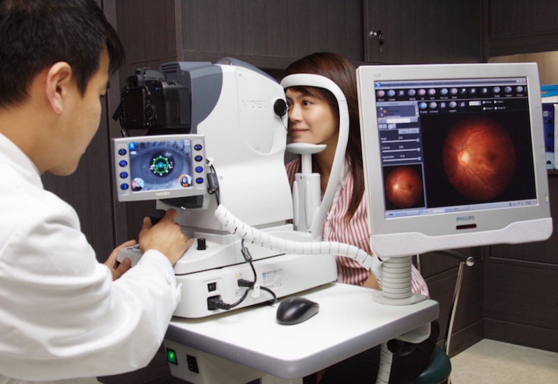
What is retinal imaging?
Retinal Imaging is a high definition, digital scan of the back of your eyes (i.e. retina). The image can show important information about your retina (e.g. optic nerve, retinal blood vessels and the area where you do your detail focusing – macula) to accurately determine the health of your eyes.
Why would I need imaging?
Many eye disease that can cause vision problems or damage in long term often have little or no symptoms at early stage, retinal imaging is very useful in early detection of disease such as macular degeneration, diabetes and glaucoma. Even for people who have no signs of eye disease, it is highly recommended as a baseline record and giving us a better way to detect changes to your eye health that need monitoring or further management. Retinal Imaging is fast and non invasive, it can give us an insight of your general health and can highlight issues with blood pressure or other conditions.What is Optical Coherence Tomography (OCT)*?
Optical Coherence Tomography (OCT) is a non-invasive imaging test. OCT uses light waves to take cross-section pictures of your retina. This measurements help to diagnosis and provide treatment guidance for glaucoma and disease of the retina e.g. age-related macular degeneration (AMD) and diabetic eye disease.*Our clinic uses fundus retinal photography as standard equipment, and OCT is usually performed after general eye examinations. If you'd like to have an OCT for more in-depth retinal health assessment /ARIA to assess the risk of stroke, we will schedule a 2nd appointment specially for these test.
